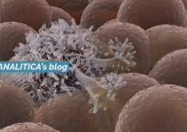
What are Spheroids? They are cell aggregates formed as a result of cell-cell adhesion
Spheroids are considered as the gold standard among the 3D in vitro models. Spheroids are cell aggregates formed as a result of cell-cell adhesion and this model is able to recapitulate cell-cell interaction as well as cell spatial arrangement.
How to produce spheroids?
Different scaffold-free methods promote the self-aggregation of cells in spheroids, among them:

- Ultra-low adhesion plates (Fig.a) are often made of coated polystyrene, that causes cells in suspension to aggregate into spheroids. Because of their larger volume, ultra-low adhesion plates are well suited for multicellular cultures, but does not allow control over spheroid size and uniformity.
- In the hanging drop technique (Fig.b), droplets of cell suspension are pipetted onto the underside of an adherent tissue culture lid, the tray is subsequently inverted and gravity induces spontaneous cell aggregation into spheroids at the bottom of the drop, at the liquid–air interface. Spheroids are tightly packed and may represent tumor layers near to a capillary. Although the spheroid size can be controlled by the initial number of cells suspended in the drop, this procedure is highly laborious.
- Magnetic levitation (Fig.c) employs magnetic nanoparticles that act as patterning agents to guide self-assembly of cells into spheroids under magnetic forces. Semi-confluent adherent cells are preloaded with magnetic nanoparticles then, the application of a magnetic field promotes self-aggregation into spheroids within few hours.
- Agitation-based approaches (Fig.d) for the production of 3D spheroids can be divided into spinner flask bioreactors and rotational culture systems. In both approaches, the cell suspension is placed into a container that is kept in motion, so cells do not adhere to the container walls, but instead form cell–cell interactions.
Pro and cons of spheroids
Spheroids have the ability to reproduce the architecture and metabolism of their tissue of origin. Moreover, they show a relative ease of self-assembly, the possibility to co-culture cancer and stromal cells (thus the possibility to mimic cancer cellular heterogeneity) and reproducibility. However, there are some limitations: not all tumor cell lines form 3D spheroids, this system exhibits a low percentage of some tissue components and this type of culture is difficult to maintain over long time periods.
Here we have shared an overview of the spheroid model, highlighting the capability to recapitulate physiological characteristics of tumors. However, the disadvantages associated with spheroids have led to the development of 3D cancer models with more structural complexity and stability.
Soon we will talk more about scaffold-based models. Stay tuned!
Author: Dr. Bianca Bazzolo
Bibliography:
- Ferreira, L. P., V. M. Gaspar, and J. F. Mano. 2018. “Design of Spherically Structured 3D in Vitro Tumor Models -Advances and Prospects.” Acta Biomaterialia 75:11–34.
- Langhans, Sigrid A. 2018. “Three-Dimensional in Vitro Cell Culture Models in Drug Discovery and Drug Repositioning.” Frontiers in Pharmacology 9(January):1–14.


